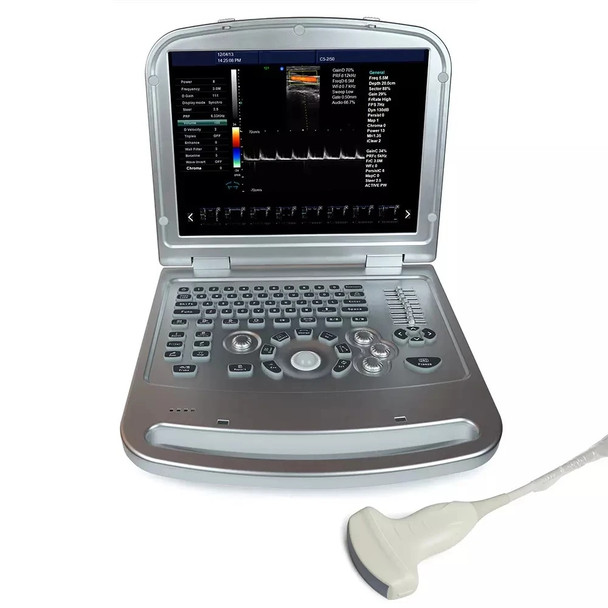
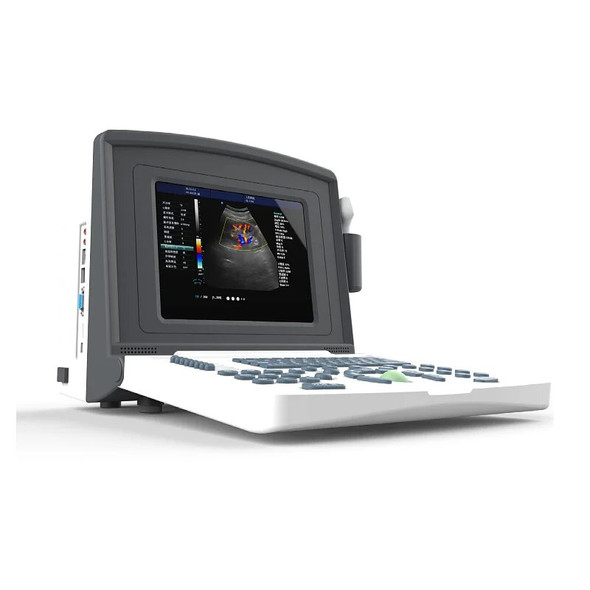
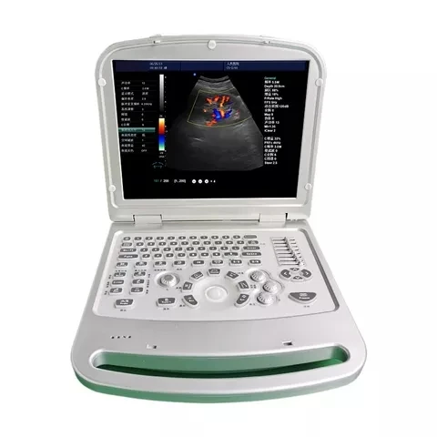
Description


Application:Abdominal, Vascular, Breast, Obstetrics, Superficial Structure, Musculoskeletal, Urology, Pediatrics/Neonatal, Small Organs, Heart, etc
Features:
Image Storage: ≧ 500 Frames
Channels : 64
Display Depth: ≧300mm
Scanning Model: Electronic Linear Array, Electronic Convex Array
LCD Size: 15 Inch
Gray Scale: 256 Levels
Image Flip: Up/ Down, Left/ Right
Image Processing: Image Smoothing/ Sharpening, Tissue Harmonic, Histogram, DR, Gamma Correction, Pseudo-Color
Measurement: Freehand, Ellipse, Distance for Perimeter, Area Volume, Heart, G.A, EDD, BPD-FW, FL, AC, HC, CRL, AD, GS, LMP
Character Display: Date, Clock, Name, PID, Age, Sex, Hospital Name, Doctor
Notation
Full-screen Character Editor, Body Mark, Position Indications
Report: One-click generation
Scanning Mode: B, B/B, 4B, B/M, M, B+C, B+D, B+C+D, PDI, CF, PW
Scanning Angle: Adjustable
Cine Loop: 200~1000 frames
External Display: PAL, VGA
USB Ports: 2
Power Consumption MAX: 100VA
Product Size: 365×385×80mm
Carton Size: 680×460×210mm
N.W./ G.W.: 10kg/ 11kg
Main technical specifications and system overview:
1. Overview of host system performance
*1.1 Operating system: Windows XP operating system, switch between Chinese/English menus
1.2 Ultra-wide band fully digital beam former: dynamic focus
1.3 Visually adjustable dynamic range
1.4 Two-dimensional gray scale imaging unit, gray scale ≥256, with excellent fine and contrast resolution and full field uniformity
1.5 Color Doppler Ultrasound Diagnostic Unit
1.6 Spectral Doppler Display and Analysis Unit
1.7 Doppler energy map, directional energy map
1.8 Tissue harmonic imaging
2. Measurement and Analysis (B-mode, M-mode, Spectral Doppler, Color Doppler)
2.1 Application Measurements
2.2 Doppler blood flow measurement and analysis
2.3 Urology Measurement and Analysis
2.4 Abdominal measurement and analysis
2.5 Small organ measurement and analysis
2.6 Obstetrics and Gynecology Measurement and Analysis
2.7 Bone Measurement and Analysis
2.8 Measurement and analysis of peripheral blood vessels
2.9 Cardiac Measurements and Analysis
3. Image storage and (movie) playback replay unit
4. Input/output signal
VGA, VIDEO, USB interface
5. Recording device:
4. Technical parameters and requirements
1. General functions of the system
1.1 Monitor: 15-inch high-resolution color LCD monitor, no flicker, can be up, down, left or right
rotate.
1.2 Operation keyboard: Silicone button, flexible and convenient operation.
1.3 Probe interface ≥ 1, fully activated, interoperable
2. Probe Specifications
*2.1 Probe frequency: ultra-wideband probe, imaging frequency range 2.0MHz—12.00MHz
2.2 Both 2D and Doppler (B/D): Linear B/PWD; Convex B/PWD
2.3 Puncture guide: optional probe can be equipped with puncture guide device (optional)
3. Two-dimensional imaging main parameters:
3.1 Scan
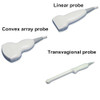
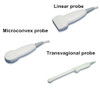
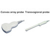
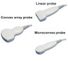
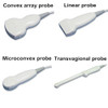
Abdominal convex array
Superficial line array
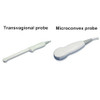
Transaginal probe
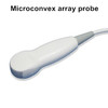
Microconvex arary probe
*3.2 Maximum display depth: ≥30cm
3.3 Real-time ZOOM zoom function
3.4 Beam deflection of linear array probe: ≥10°
3.5 Speed of sound focusing: ≥4 focal points, the position is continuously adjustable
3.6 Playback reproduction: large-capacity movie playback
3.7 Ultrasonic output power: Visually adjustable
3.8 Digital Beamformer: Continuous Dynamic Focus, Variable Aperture and Dynamic Zoom
3.9 Preset conditions: For different inspection organs, preset the inspection conditions to optimize the image to reduce the adjustment during operation
*3.10 Image optimization technology: Visually adjustable ≥6 files
3.11 Gain adjustment: 8-segment TGC adjustment
3.12 M-type scanning speed ≥3 gears adjustable
3.13 B/M layout can be adjusted visually
4. Pulse Doppler
4.1 Deflection angle: -2.5—10 four-stage adjustable
4.2 Pulse repetition frequency: 1.82KHZ-7.52KHZ adjustable
4.3 Spectrum Inversion
4.4 Triple synchronization
4.5 Wall filter ≥3 levels adjustable
4.6 Spectral pseudo-color ≥ 6 adjustable
4.7 Spectrum envelope function: multiple modes of real-time spectrum envelope are optional, and the system automatically analyzes and displays: PSV, EDV, AT, DT, RI, PI, SD, HR and other data
4.8 Edge enhancement ≥ 8 adjustable
4.7 Baseline ≥ 7 adjustable
5. Color Doppler
5.1 Doppler probe and frequency: Linear array: PWD; Convex array: PWD
All of the above probes have two or more Doppler imaging operating frequencies and are independently adjustable
5.2 Wall filter: ≥3 grades, continuously adjustable
5.3 Display position adjustment: line scan Interested image range: -10°~+10°
6. Ultrasound image and medical record management system
6.1 The compression ratio of the dynamic image can be adjusted, and it can be directly executed on an ordinary PC without special software.
Observe the image
6.2 Dynamic image acquisition and storage, quick “one-finger” image review
6.3 Images can be embedded in medical record reports and edited
6.4 Dedicated measurements and analyses
6.5 Fetal Growth Curve
6.6 Storing images and documents: U disk
6.7 Report Storage, Retrieval, Statistics
6.8 Full digital high-capacity hard disk ≥120G
Package list:
Machine main unit 1pc
probes depend on your requirements
Warranty card 1pc
User's manual 1pc
Power line 1pc
Optional:
convex array probe
high frequency linear probe
transvagional probe
micro-convex array probe
Trolley cart




Our company is established in 2018 located in Chengdu. We are dedicated to the development and production of medical devices. Our products are widely recognized and trusted by users and can meet continuously changing economic and social needs.We welcome new and old customers contact us for future business relationships and mutual success!To learn more about what we can do for you, contact us at any time. We are looking forward to establishing a good and long-term business relationship with you.


If you are satisfy with our product, kindly give us 5 stars


